
Immune-organoid model systems may be the next frontier in organoid technology. By creating controlled, patient-specific micro-environments, these models provide critical mechanistic insights into immune-driven diseases and offer a high-fidelity platform for evaluating the safety and efficacy of next-generation immunotherapies.
The intricate relationship between the immune system and the body’s epithelial barriers is fundamental to health, but its complexity has hindered mechanistic study. Traditional cultures lack physiological relevance, while animal models fail to fully capture the nuances of human immunology, creating a critical gap in understanding immune-driven diseases.
In a recent review in Cell Reports, PhD student and first author Marius Harter (IHB and University Paris-Saclay) and co-authors Timothy Recaldin (Roche pRED), Nikolche Gjorevski (IHB) break down how these co-culture systems can reconstruct key phases of a functional immune response. For instance, organoids can mimic the initial “detection” phase by recruiting immune cells like neutrophils in response to microbial cues. By incorporating specific effector cells such as intraepithelial lymphocytes (IELs), researchers can model the targeted “deployment” phase and map precise signaling pathways. The models also shed light on the critical “resolution” phase, where regulatory cells work to restore tissue homeostasis.
These systems have direct translational applications, from modeling T cell-driven epithelial damage in inflammatory bowel disease (IBD) to testing the efficacy and safety of cancer immunotherapies using patient-derived tumoroids. The authors conclude by highlighting the next frontier: integrating these co-cultures with bioengineering approaches like organs-on-chips to create vascularized models that replicate the entire organ immunity cycle, opening new avenues for understanding and treating human immunological disorders.
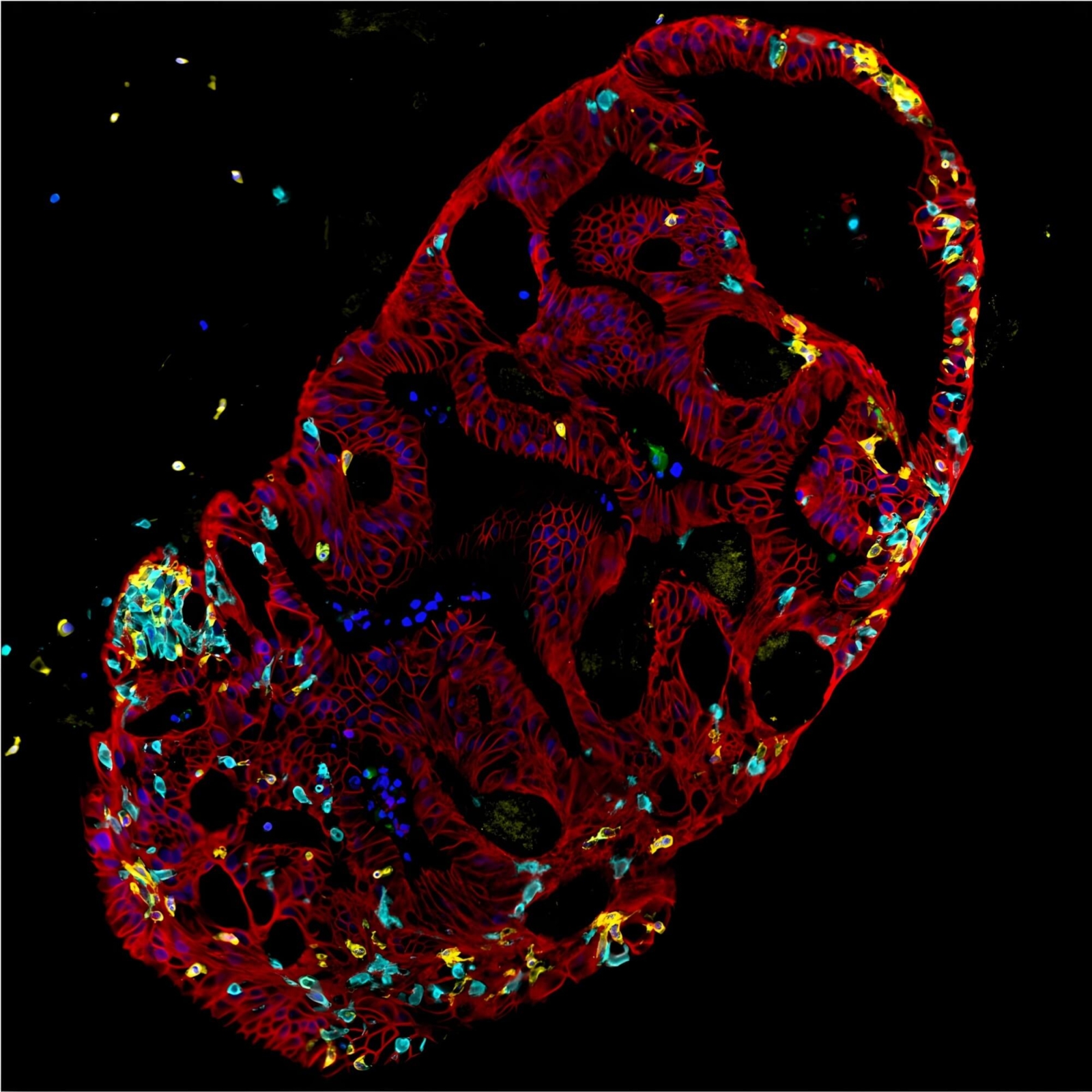
Immune-epithelial cell interactions under the microscope: multiplex-immunofluorescent image of tissue-resident memory T cells (CD4+, yellow; CD8+ ,cyan). T cells integrating and surveilling the epithelium of a donor-matched intestinal organoid (E-Cadherin+, red; Nuclei, blue). Credit: Marius Harter.
Available open access: Harter MF, Recaldin T, Gjorevski N. Organoids as models of immune-organ interaction. Cell Reports 2025. DOI: 10.1016/j.celrep.2025.116214
Read the full paper: https://www.sciencedirect.com/science/article/pii/S2211124725009854